Colon Drawing
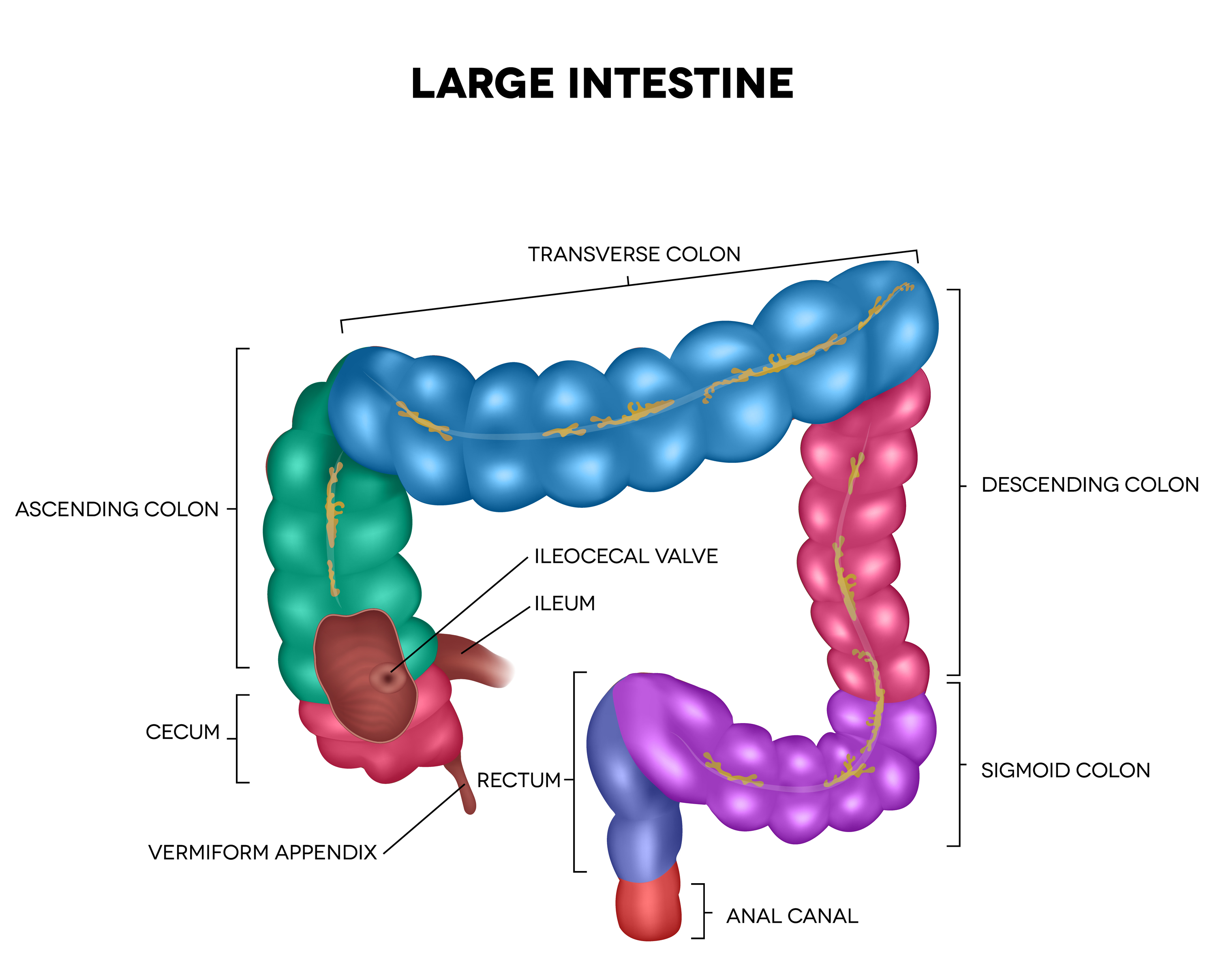
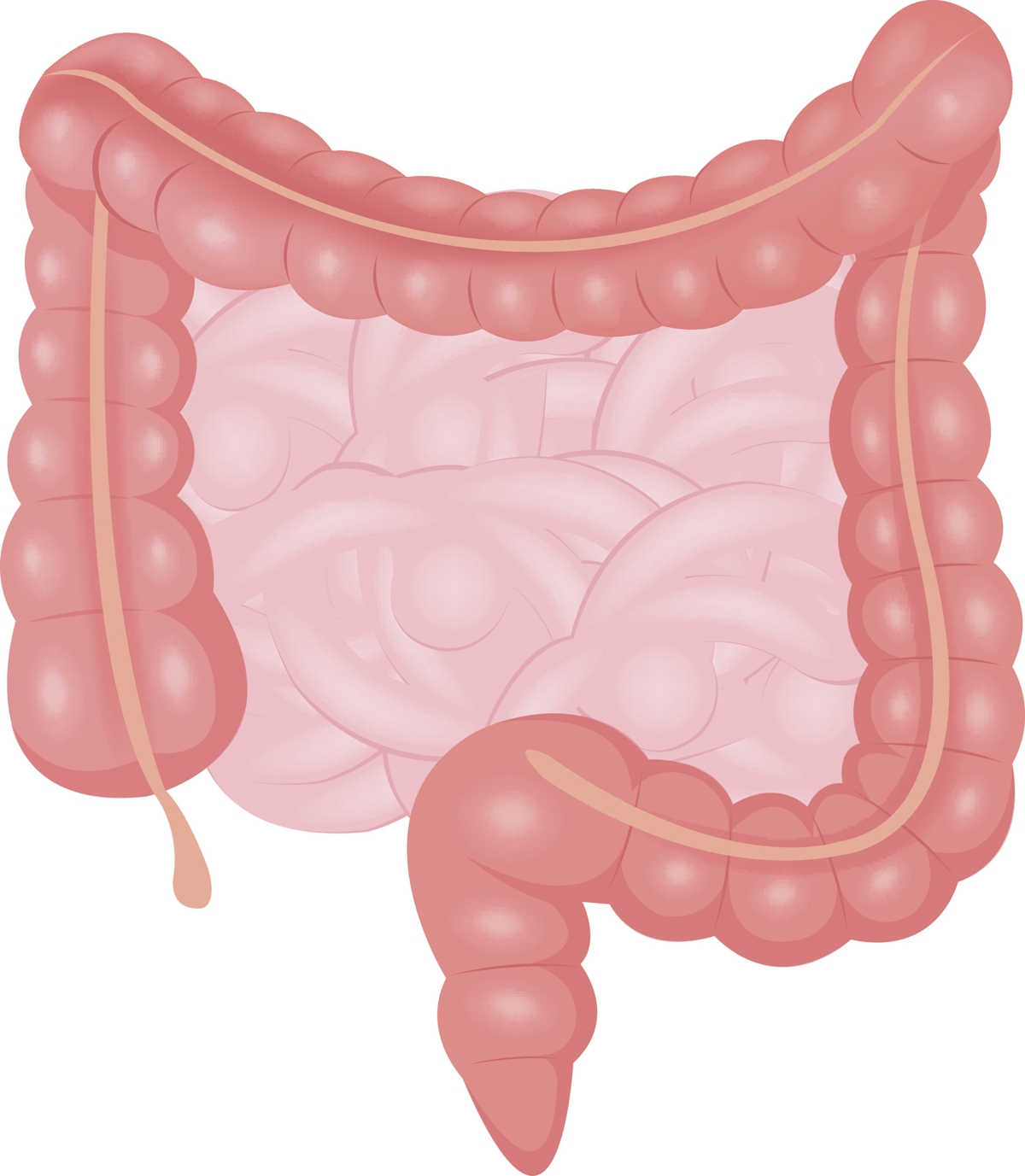
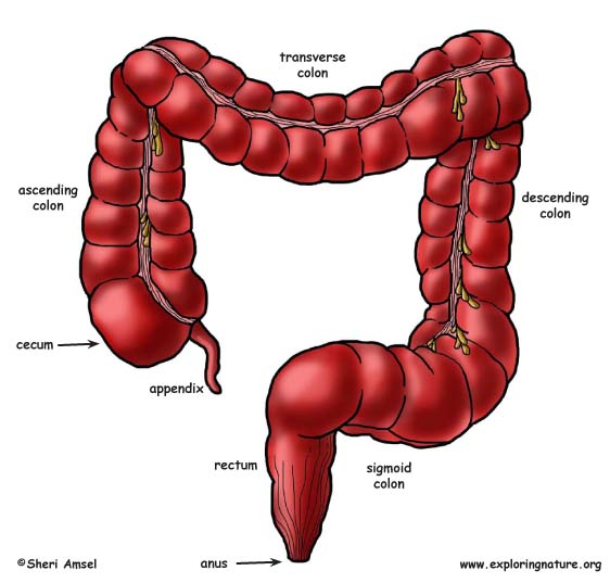
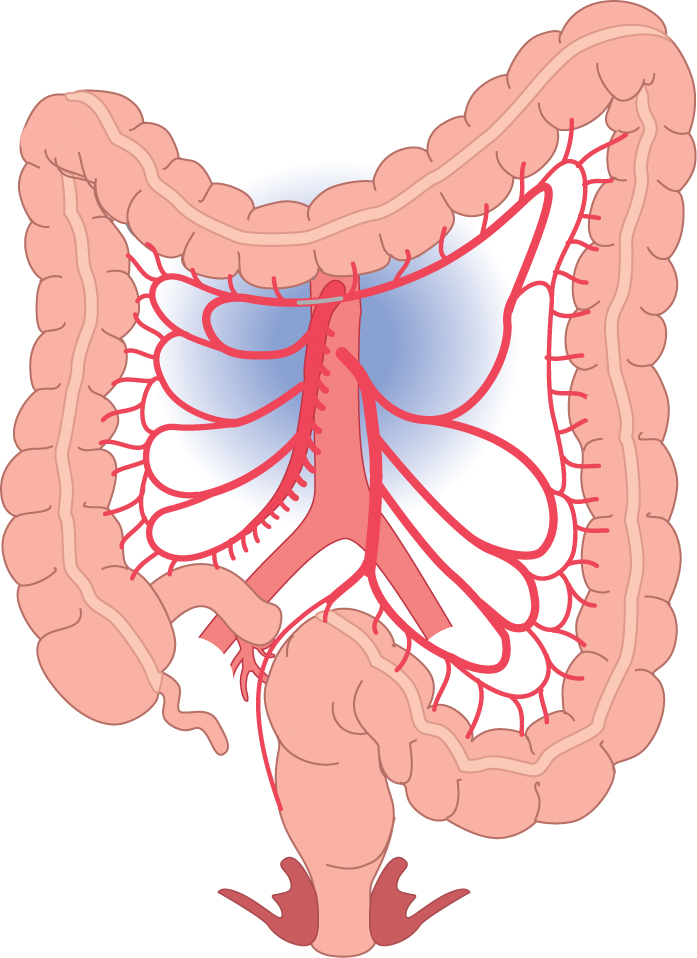
Colon Drawing - Drawing of colon stock illustrations. The rectum is a hollow muscular tube about 8 inches (20 cm) in length and 2.5 inches in diameter at its widest point. Web the colon may be subdivided into four parts: Also shown is the small intestine. The colon is made of several sections. Copyright 2021 american society of clinical oncology. Web how to draw a model of the digestive system. The ascending colon lies secondary retroperitoneally on the right side of the abdominal cavity and moves towards the right colic flexure at the bottom side of the liver. Colon, also called the large intestine; Drawing of the gi tract, with the esophagus, stomach, small intestine, duodenum, jejunum, ileum, large intestine, cecum, colon, rectum, and anus labeled. The rectum is a hollow muscular tube about 8 inches (20 cm) in length and 2.5 inches in diameter at its widest point. March 30, 2024 fact checked. This specimen clearly shows the outer layer of the muscularis externa as three distinct bundles (taeniae coli). Brain, kidney, heart, liver, stomach. Ascending, transverse, descending and sigmoid colon. The colon reabsorbs water from digested food and concentrates waste material called stool. Web how to draw a model of the digestive system. It extends from the inferior end of the sigmoid colon along the anterior surface of the sacrum and coccyx in. Web parts of the colon; The colon is made of several sections. Web colon anatomy (with small intestine label) description: Web the colon is part of the large intestine, and it absorbs water and nutrients from food. Identifying features of a colon histology slide. Lamina propria of the colon slide. Web how to draw a model of the digestive system. The large intestine turns food waste into stool and passes it from the body when you poop. Breast cancer) to search by that exact term or phrase, no variations. Drawing of colon stock illustrations. Drawing shows parts of the proximal colon, including the transverse colon, ascending colon, and cecum. Search by image or video. View drawing of colon videos. This specimen clearly shows the outer layer of the muscularis externa as three distinct bundles (taeniae coli). You will find a drawing of the main body parts affected by colorectal cancer. Lamina propria of the colon slide. Approved by the cancer.net editorial board, 09/2023. Web table of contents. Lamina propria of the colon slide. Identification points of a colon microscope slide. Also shown is the small intestine. Breast cancer) to search by that exact term or phrase, no variations. Epithelium lining of the colon. Image information and view/download options. Colon, also called the large intestine; The large intestine begins at the cecum. Drawing shows the cecum, ascending colon, transverse colon, descending colon, sigmoid colon, rectum, and anal canal. Web parts of the colon. The colon is the largest part of the large intestine, extending from the cecum to the rectum. Drawing of the front of the abdomen that shows the four sections of the colon: Search by image or video. Drawing of the gi tract, with the esophagus, stomach, small intestine, duodenum, jejunum, ileum, large intestine, cecum, colon,. Search by image or video. The ileum (small intestine) ends where it connects to the cecum at the ileocecal junction. Use the search page for more options. Identifying features of a colon histology slide. Stool travels through the colon to the latter part of the colon, the rectum. Stool travels through the colon to the latter part of the colon, the rectum. Drawing shows the ascending colon, cecum, transverse colon, descending colon, sigmoid colon, and rectum. Drawing of the colon, ileum, stoma of the ileum, rectum, and anus within an outline of the human body. The proximal colon includes the transverse colon, ascending colon, and the cecum. Web. Colon, also called the large intestine; Copyright 2021 american society of clinical oncology. Drawing shows the cecum, ascending colon, transverse colon, descending colon, sigmoid colon, rectum, and anal canal. The colon is divided into four parts: Drawing shows the ascending colon, cecum, transverse colon, descending colon, sigmoid colon, and rectum. Web parts of the colon. Web the colon may be subdivided into four parts: Drawing shows parts of the proximal colon, including the transverse colon, ascending colon, and cecum. Drawing of the gi tract, with the esophagus, stomach, small intestine, duodenum, jejunum, ileum, large intestine, cecum, colon, rectum, and anus labeled. Web image collection gallery list. Web how to draw a model of the digestive system. Use the menu to see other pages. Drawing of colon stock illustrations. The colon reabsorbs water from digested food and concentrates waste material called stool. It’s all one, long tube that continues from the small intestine as food nears the end of its journey through your digestive system. It is 5 feet long and its function is to reabsorb water from digested food and concentrate solid waste material, known as stool.Structure And Function Of The Large Intestine Anatomy System Stock
Large Intestine Human Anatomy Stock Illustration Download Image Now
Large Intestine Detailed Illustration gutCARE

Colon, Drawing Stock Image C021/3991 Science Photo Library

Anatomy of the caecum, appendix and colon Surgery Oxford
Large Intestine Anatomy Bodytomy
Large Intestine Colon Sections Labeled In Male Abdominal Anatomy Stock
Large Intestine (Colon)
Slagter Drawing Colon and rectum anatomy and arteries no labels

Colon, illustration Stock Image F012/7551 Science Photo Library
Web Drawing Of The Colon With The Ascending Colon, Transverse Colon, Descending Colon, And Sigmoid Colon Labeled.
This Specimen Clearly Shows The Outer Layer Of The Muscularis Externa As Three Distinct Bundles (Taeniae Coli).
The Ileum (Small Intestine) Ends Where It Connects To The Cecum At The Ileocecal Junction.
Lamina Propria Of The Colon Slide.
Related Post:


