Drawing Of Sinuses
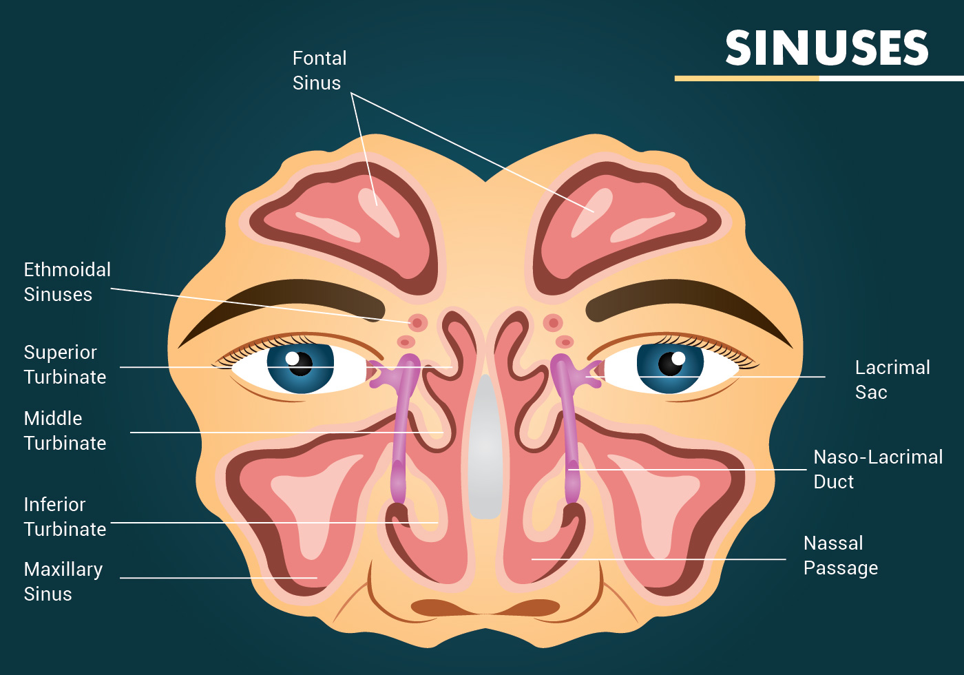
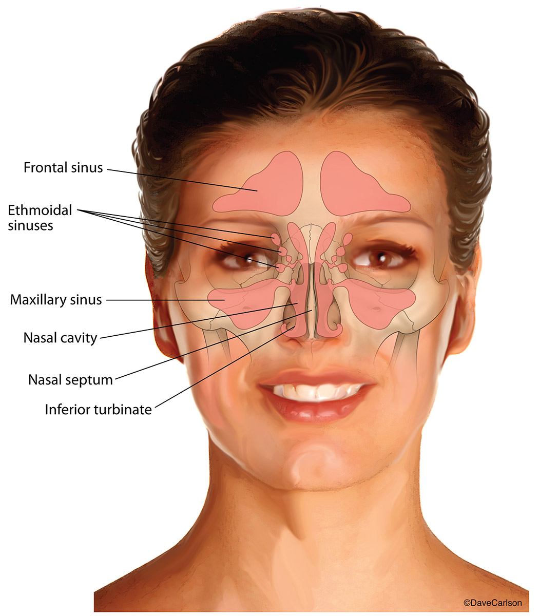
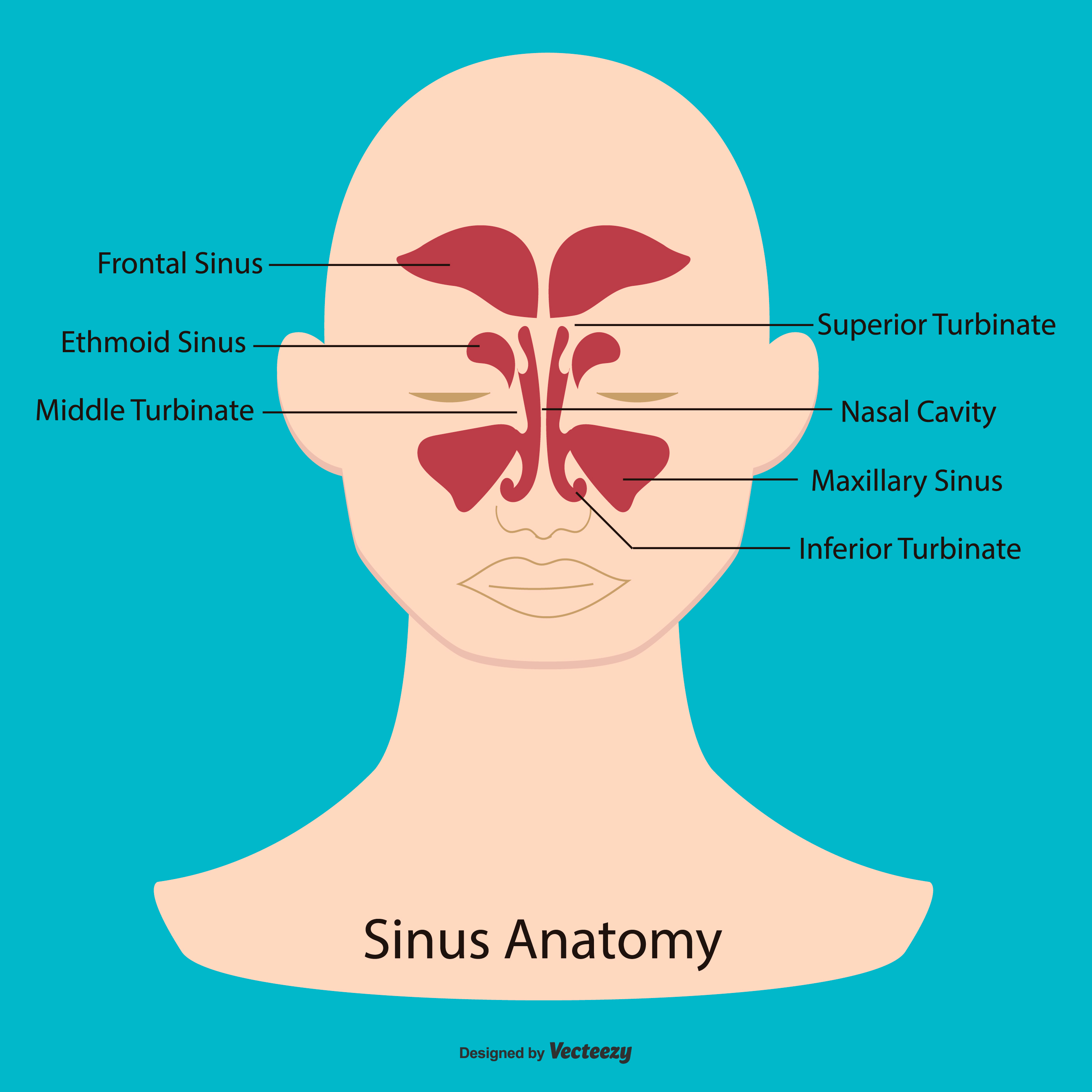
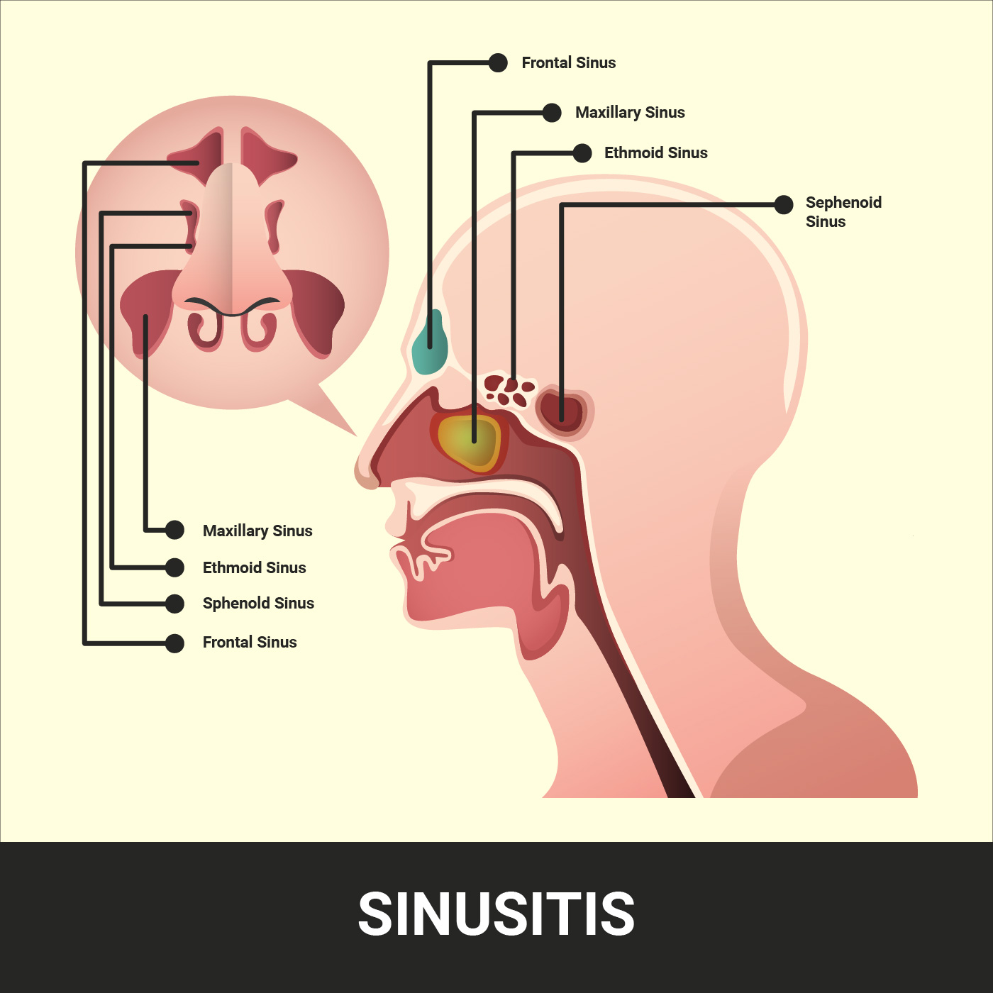
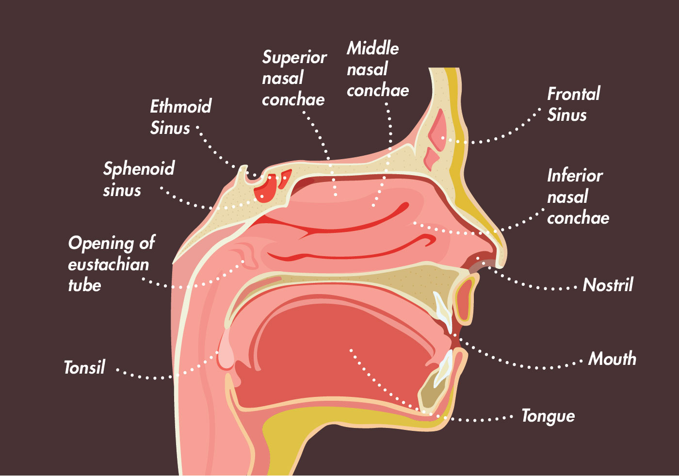
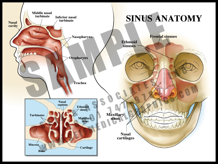
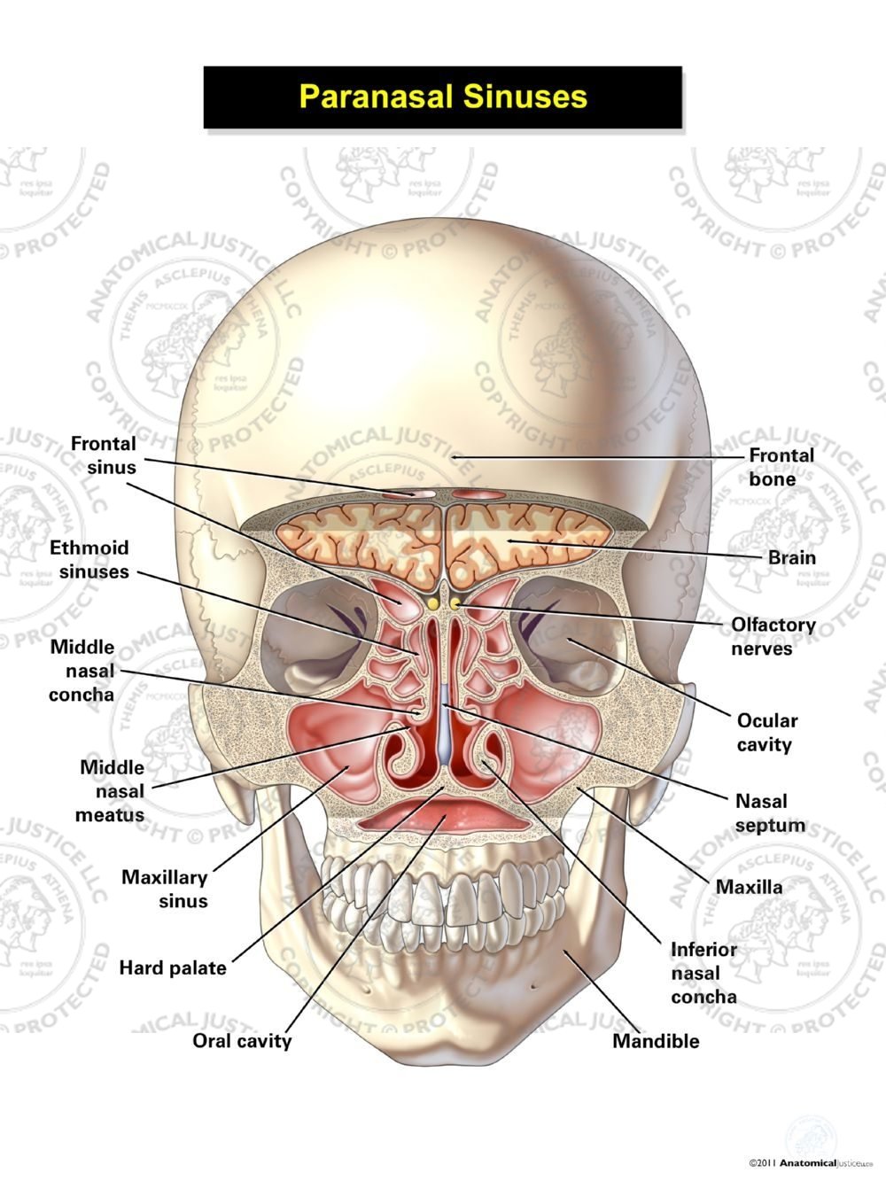
Drawing Of Sinuses - The maxillary sinuses are behind the cheeks. The external nasal anatomy is quite simple. Web it also can whisk away bacteria, thin mucus, and cut down on postnasal drip. The sphenoid sinuses are located behind the ethmoid sinuses. Learn this topic now at kenhub! Web the ethmoidal cells (sinuses) the prime function of the paranasal sinuses is to protect the organism, mostly by humidifying the inhaled air and facilitating the immune response of the respiratory system. We also go over sinusitis signs and care. Web anatomy of the sinuses. Next, we present the anatomy of the nasal cavity, nasal septum, and lateral nasal wall with their blood supply and innervation. Web browse 20 sinuses diagram photos and images available, or start a new search to explore more photos and images. Web nasal anatomy skull base anatomy. Next, we present the anatomy of the nasal cavity, nasal septum, and lateral nasal wall with their blood supply and innervation. Web the ethmoidal cells (sinuses) the prime function of the paranasal sinuses is to protect the organism, mostly by humidifying the inhaled air and facilitating the immune response of the respiratory system. The. Web there are 4 pairs of the paranasal sinuses: Web it also can whisk away bacteria, thin mucus, and cut down on postnasal drip. The app incorporates 3d schematic models of the following: Web in this chapter we first review embryology, then review the surface anatomy of the external nose, the nasal framework, and the nasal musculature, along with their. These pockets of air, located in the bones of our faces, not only strengthen the skull but also filter the air that we breathe. Web it is attached to the lateral nasal wall posteriorly just above the inferior turbinate but behind the maxillary, or cheek, sinus. Whereas it would normally be impossible to drain a “dead end” cavity like the. We also go over sinusitis signs and care. Openstax college, cc by 3.0, via wikimedia commons. Anatomy and physiology serve as the basis for understanding diseases of the nose and sinuses, and we have provided brief summaries of this to help with your foundation of understanding. Web browse 20 sinuses diagram photos and images available, or start a new search. Web browse 20 sinuses diagram photos and images available, or start a new search to explore more photos and images. The nasal cavity and pharynx (throat) are also shown. The apex of the sinus extends into the zygomatic process of the maxilla, and the lateral wall of the nose forms the base. In the following section, you will find information. The frontal sinus, maxillary sinus, and anterior ethmoid sinus cells drain beneath the middle turbinate into the middle meatus. Web it also can whisk away bacteria, thin mucus, and cut down on postnasal drip. Web anatomy of the sinuses. Anatomy of the paranasal sinuses (spaces between the bones around the nose). The external nasal anatomy is quite simple. Whereas it would normally be impossible to drain a “dead end” cavity like the sinuses, jal neti achieves this ingeniously and simply. Web browse 237 sinuses anatomy photos and images available, or search for human sinuses anatomy to find more great photos and pictures. Web nasal anatomy skull base anatomy. The external nasal anatomy is quite simple. Learn this topic. Learn this topic now at kenhub! Sphenoid bone and occipital bone, human skull, victorian anatomical drawing. To understand the work our sinuses do, we must examine sinus anatomy. These pockets of air, located in the bones of our faces, not only strengthen the skull but also filter the air that we breathe. Most of the time our sinuses perform an. The apex of the sinus extends into the zygomatic process of the maxilla, and the lateral wall of the nose forms the base. Web browse 237 sinuses anatomy photos and images available, or search for human sinuses anatomy to find more great photos and pictures. In the following section, you will find information pertaining to the anatomy of the nasal. Superiorly, it inserts along the lateral nasal wall and skull base. Sphenoid bone and occipital bone, human skull, victorian anatomical drawing. Web in this chapter we first review embryology, then review the surface anatomy of the external nose, the nasal framework, and the nasal musculature, along with their blood supply and innervation. Drawing shows front and side views of the. Web anatomy atlas of the nasal cavity: Fully labeled illustrations and diagrams of the nose and paranasal sinuses (external nose, nasal cartilages, nasal septum, nasal concha and meatus, bones of the nasal cavity and vessels and nerves). We also go over sinusitis signs and care. Web the ethmoidal cells (sinuses) the prime function of the paranasal sinuses is to protect the organism, mostly by humidifying the inhaled air and facilitating the immune response of the respiratory system. There are several sets of sinuses in the head. Web in this chapter we first review embryology, then review the surface anatomy of the external nose, the nasal framework, and the nasal musculature, along with their blood supply and innervation. Web it is attached to the lateral nasal wall posteriorly just above the inferior turbinate but behind the maxillary, or cheek, sinus. The nasal cavity and pharynx (throat) are also shown. Web browse 20 sinuses diagram photos and images available, or start a new search to explore more photos and images. Web it also can whisk away bacteria, thin mucus, and cut down on postnasal drip. Maxillary sinuses (the biggest) frontal sinuses. Next, we present the anatomy of the nasal cavity, nasal septum, and lateral nasal wall with their blood supply and innervation. The maxillary sinuses are behind the cheeks. Anatomy of the paranasal sinuses (spaces between the bones around the nose). Openstax college, cc by 3.0, via wikimedia commons. The apex of the sinus extends into the zygomatic process of the maxilla, and the lateral wall of the nose forms the base.
Sinus Anatomy, Illustration Stock Image C036/6312 Science Photo
Sinuses Vector Design 172613 Vector Art at Vecteezy

Structure Of Sinuses
Nasal Sinuses Carlson Stock Art
Sinus Anatomy Illustration 172412 Vector Art at Vecteezy
Sinus Vector Illustration With Detailed Information 171464 Vector Art

Sinus, Drawing Stock Image C017/1681 Science Photo Library
Sinuses Vector Design 170605 Vector Art at Vecteezy
Sinus Anatomy S&A Medical Graphics
Paranasal Sinuses Illustration Anatomical Justice
Web Anatomy Of The Sinuses.
These Pockets Of Air, Located In The Bones Of Our Faces, Not Only Strengthen The Skull But Also Filter The Air That We Breathe.
Anatomy And Physiology Serve As The Basis For Understanding Diseases Of The Nose And Sinuses, And We Have Provided Brief Summaries Of This To Help With Your Foundation Of Understanding.
To Understand The Work Our Sinuses Do, We Must Examine Sinus Anatomy.
Related Post: