Female Anatomy Drawing Labeled
Female Anatomy Drawing Labeled - The function of your external genitals are to protect the internal parts from infection and allow sperm to enter your vagina. A lot of people mistakenly use the term “vagina” to. This article looks at female body parts and their functions, and it provides an interactive. The vagina is a muscular canal lined with nerves and mucus membranes. It connects the uterus and cervix to the outside of the. Anatomy of the female reproductive system; Egg cell production, or oogenesis, begins with the primordial follicles. Web learn anatomy as you browse our collection of colorful, large and clearly labeled human body diagrams. Web zygote body is a free online 3d anatomy atlas. An female’s internal reproductive organs are the vagina, uterus, fallopian tubes, cervix, and ovary. Web zygote body is a free online 3d anatomy atlas. Web the external female genitalia are a part of the female reproductive system, and include the: For teachers, students, health professionals, or anyone interested in learning about the anatomy of the human body. Web the vulva includes the: 101 labeled illustrations of the female genital system (ovaries, uterine tubes, uterus,. Frontal view of full male and female. Web one of the best ways to consolidate your knowledge of different structures is to draw them. Web the vulva includes the: Web anatomy atlas of the female pelvis: They produce oocytes (egg cells), as well as estrogen, progesterone, and other hormones. This area provides support for the. Web the female reproductive anatomy includes both external and internal parts. Web the external female genitalia are a part of the female reproductive system, and include the: Frontal view of full male and female. Web female anatomy includes the internal and external reproductive organs. The components of the external female genitalia occupy a large part of the female perineum and collectively form what's known as the vulva. The vagina is a muscular canal lined with nerves and mucus membranes. This article provides diagrams with supporting information to help you learn about the main structures and functions. They produce oocytes (egg cells), as well as. Web the major organs of the female reproductive system include the vagina, uterus, ovaries, and fallopian tubes. The functions of these organs. Web what’s the difference between the vulva and vagina? The components of the external female genitalia occupy a large part of the female perineum and collectively form what's known as the vulva. New 3d rotate and zoom. What is the female pelvis? The functions of these organs. For teachers, students, health professionals, or anyone interested in learning about the anatomy of the human body. Web zygote body is a free online 3d anatomy atlas. A lot of people mistakenly use the term “vagina” to. Web one of the best ways to consolidate your knowledge of different structures is to draw them. The superior branch of the uterine artery supplies the body and fundus, while the inferior branch supplies the cervix. With the help of three female anatomy diagrams, flo experts outline everything you need to know. The components of the external female genitalia occupy. Anatomy of the female reproductive system; The function of your external genitals are to protect the internal parts from infection and allow sperm to enter your vagina. Web anatomy atlas of the female pelvis: Frontal view of full male and female. New 3d rotate and zoom. Old engraved illustration of the female body shape. The vagina, shown at the bottom of figure 27.9 and figure 27.10, is a muscular canal (approximately 10 cm long) that serves as the entrance to the reproductive tract.it also serves as the exit from the uterus during menses and childbirth. The uterus is supplied mainly by the uterine artery which arises. Web our labeled diagrams and quizzes on the female reproductive system are the best place to start. Ovaries are the female gonads. The superior branch of the uterine artery supplies the body and fundus, while the inferior branch supplies the cervix. This article provides diagrams with supporting information to help you learn about the main structures and functions. They produce. Ovaries are the female gonads. External structures include the mons pubis, pudendal cleft, labia majora and minora, vulva, bartholin’s gland, and the clitoris. The female reproductive organs include several key structures, such as the ovaries, uterus, vagina, and vulva. The uterus is supplied mainly by the uterine artery which arises from the internal iliac artery. For teachers, students, health professionals, or anyone interested in learning about the anatomy of the human body. Web browse 630 female anatomy diagram photos and images available, or start a new search to explore more photos and images. From male anatomy to female anatomy and every structure in between, you can trust kenhub to provide the most accurate and beautifully clear drawing reference images. Web reproductive system, female, anatomy description: Web the external female genitalia are a part of the female reproductive system, and include the: Frontal view of full male and female. Drawing shows the uterus, myometrium (muscular outer layer of the uterus), endometrium (inner lining of the uterus), ovaries, fallopian tubes, cervix, and vagina. The superior branch of the uterine artery supplies the body and fundus, while the inferior branch supplies the cervix. Although the external female genitalia are commonly referred to as the “vagina,” the vagina is just one of several organs that comprise the external female. 101 labeled illustrations of the female genital system (ovaries, uterine tubes, uterus, vagina, vulva, clitoris) and pelvic cavity (bladder, rectum, pelvic diaphragm, perineum with innervation and blood supply) Web female anatomy includes the internal and external reproductive organs. Web female anatomy includes the external genitals, or the vulva, and the internal reproductive organs.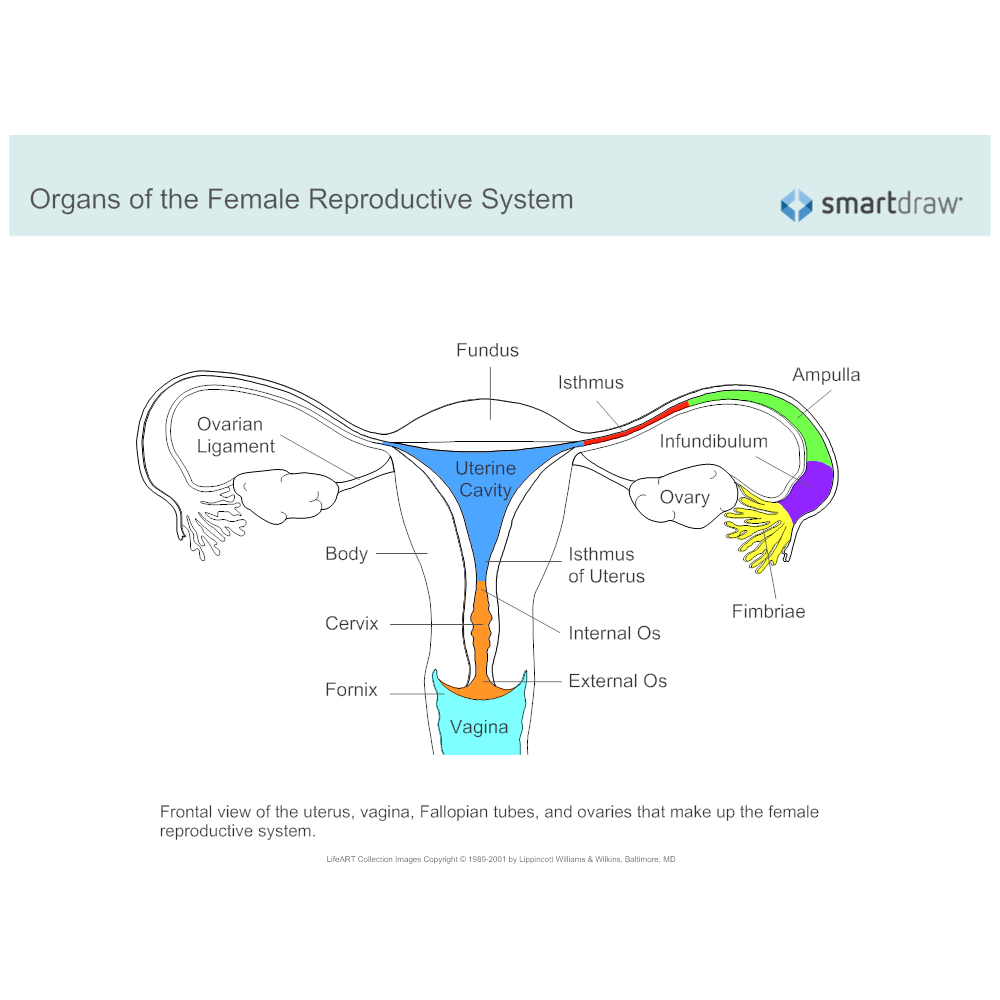
Female Reproductive System Diagram
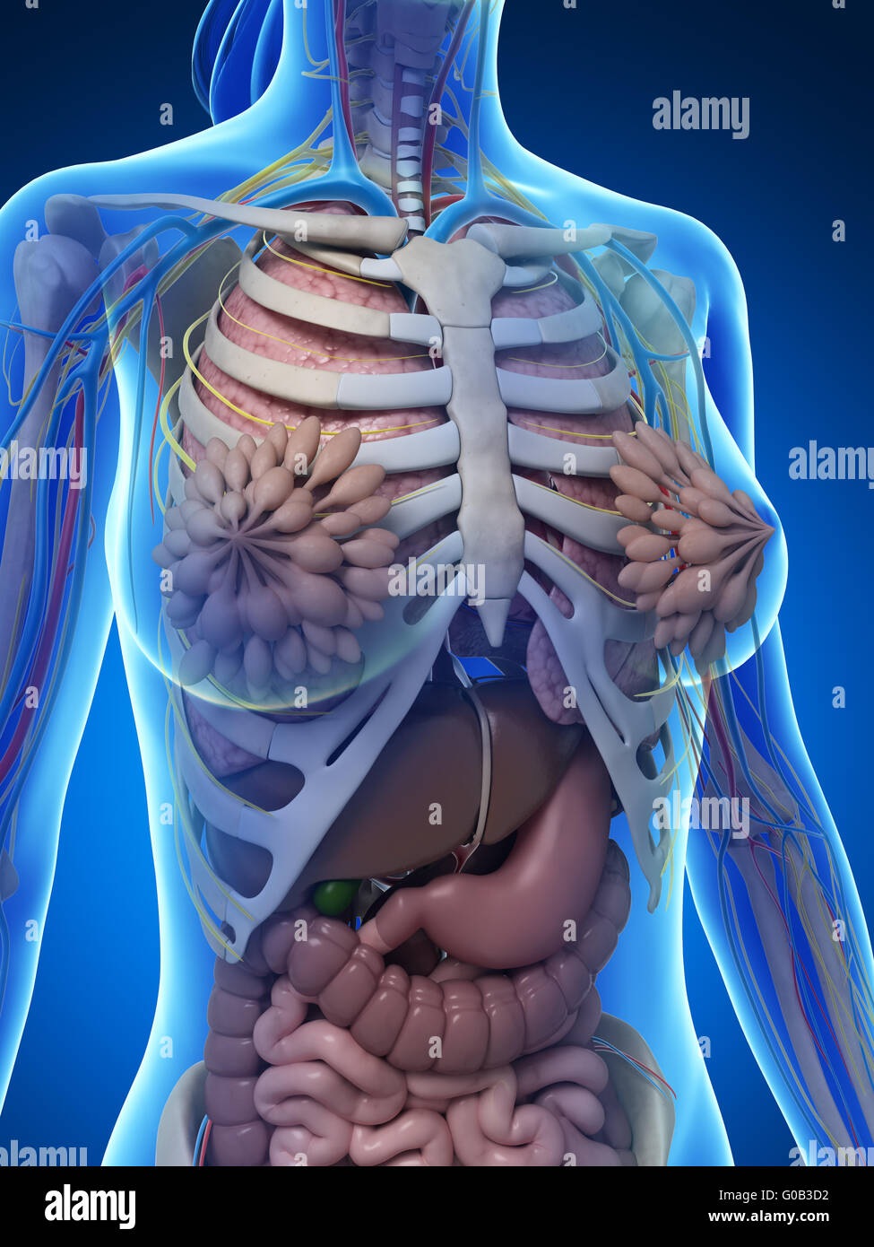
Female anatomy diagram hires stock photography and images Alamy
Female anatomy stock illustration. Illustration of human 30725871

Illustration Of Woman\'S Internal Organs Human Organs Stock Photos
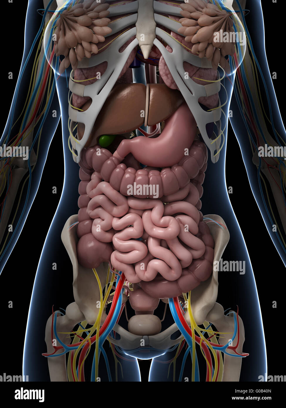
Female anatomy diagram hires stock photography and images Alamy

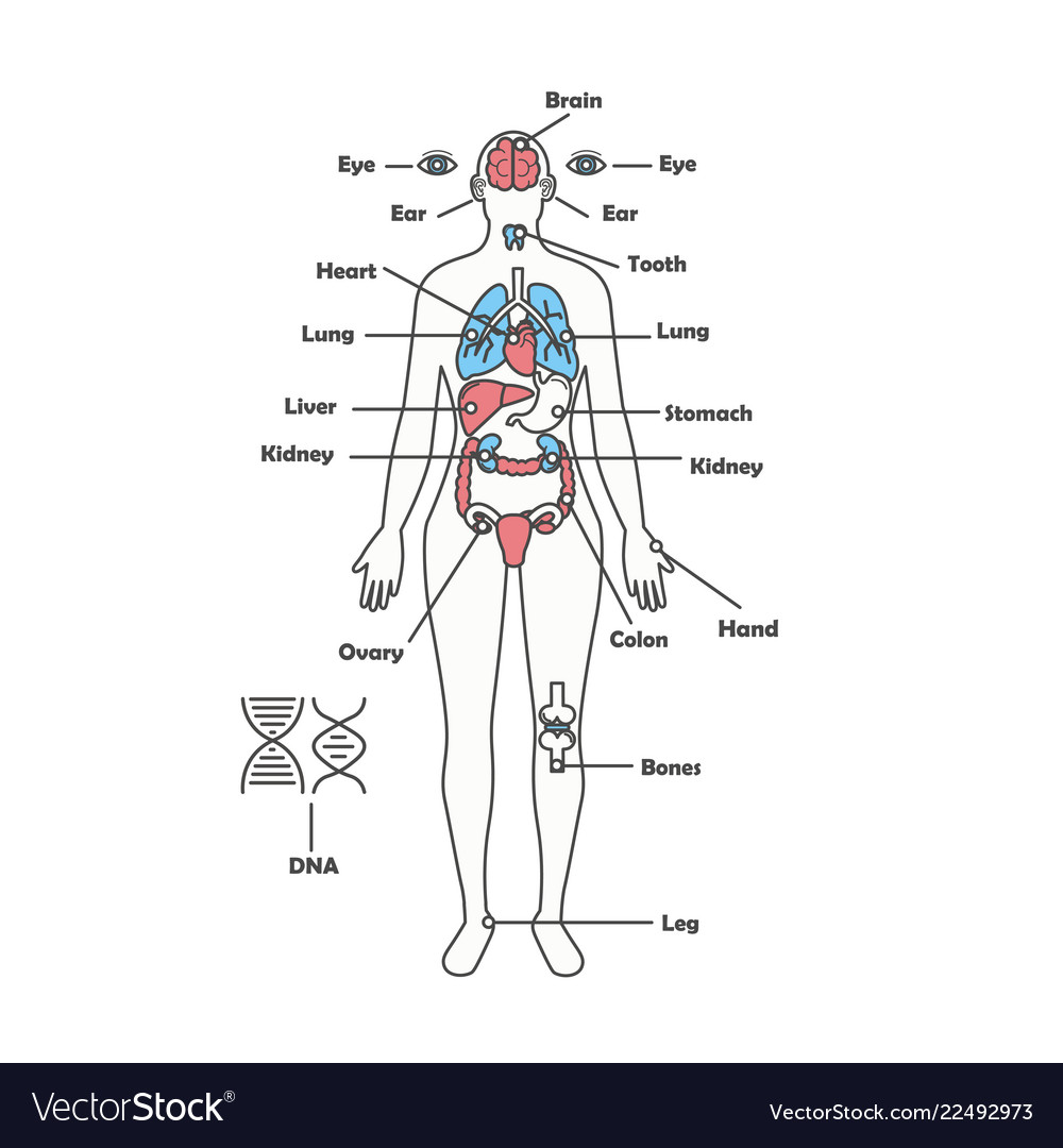
Female human anatomy body systems Royalty Free Vector Image
Female Anatomy Diagram Stock Photos & Female Anatomy Diagram Stock
Anatomy Of Female Body Organs Vertebrae Rays Lumbar Ray Spinal Spine
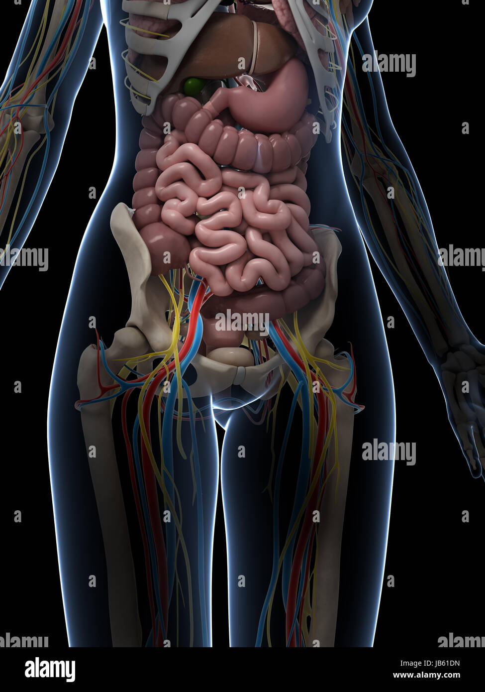
Female Anatomy Diagram Stock Photos & Female Anatomy Diagram Stock
Female Anatomy Diagram Stock Photos & Female Anatomy Diagram Stock
Although A Man Is Needed To Reproduce, It Is The Woman Who Incubates The Developing.
Web One Of The Best Ways To Consolidate Your Knowledge Of Different Structures Is To Draw Them.
The Vagina, Shown At The Bottom Of Figure 27.9 And Figure 27.10, Is A Muscular Canal (Approximately 10 Cm Long) That Serves As The Entrance To The Reproductive Tract.it Also Serves As The Exit From The Uterus During Menses And Childbirth.
Mons Pubis, Labia Majora, Labia Minora, Clitoris, Vestibule, Hymen, Vestibular Bulb And Vestibular Glands.
Related Post: