Labelled Heart Drawing
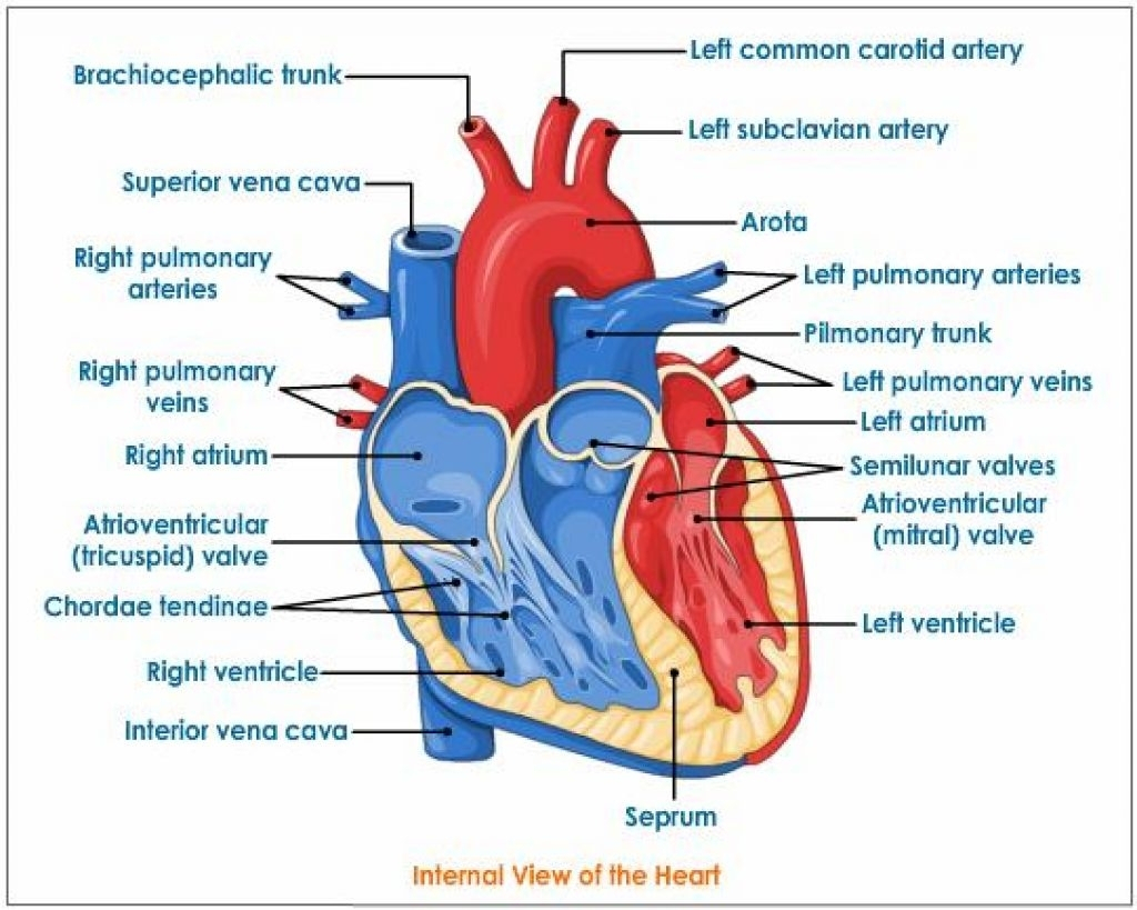
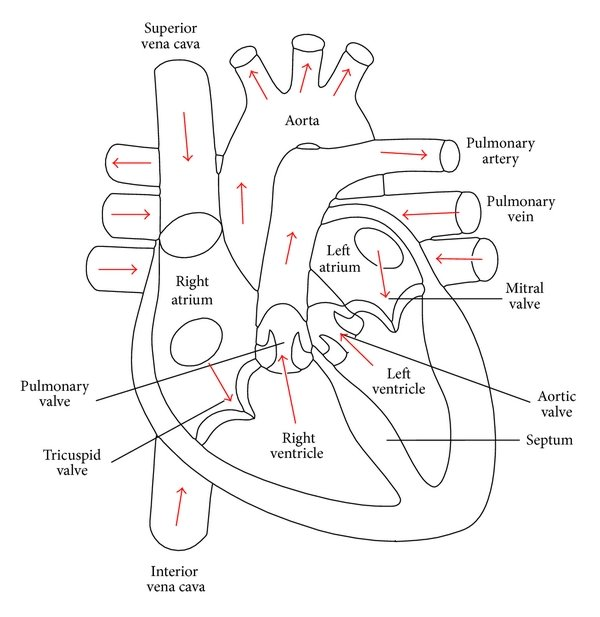
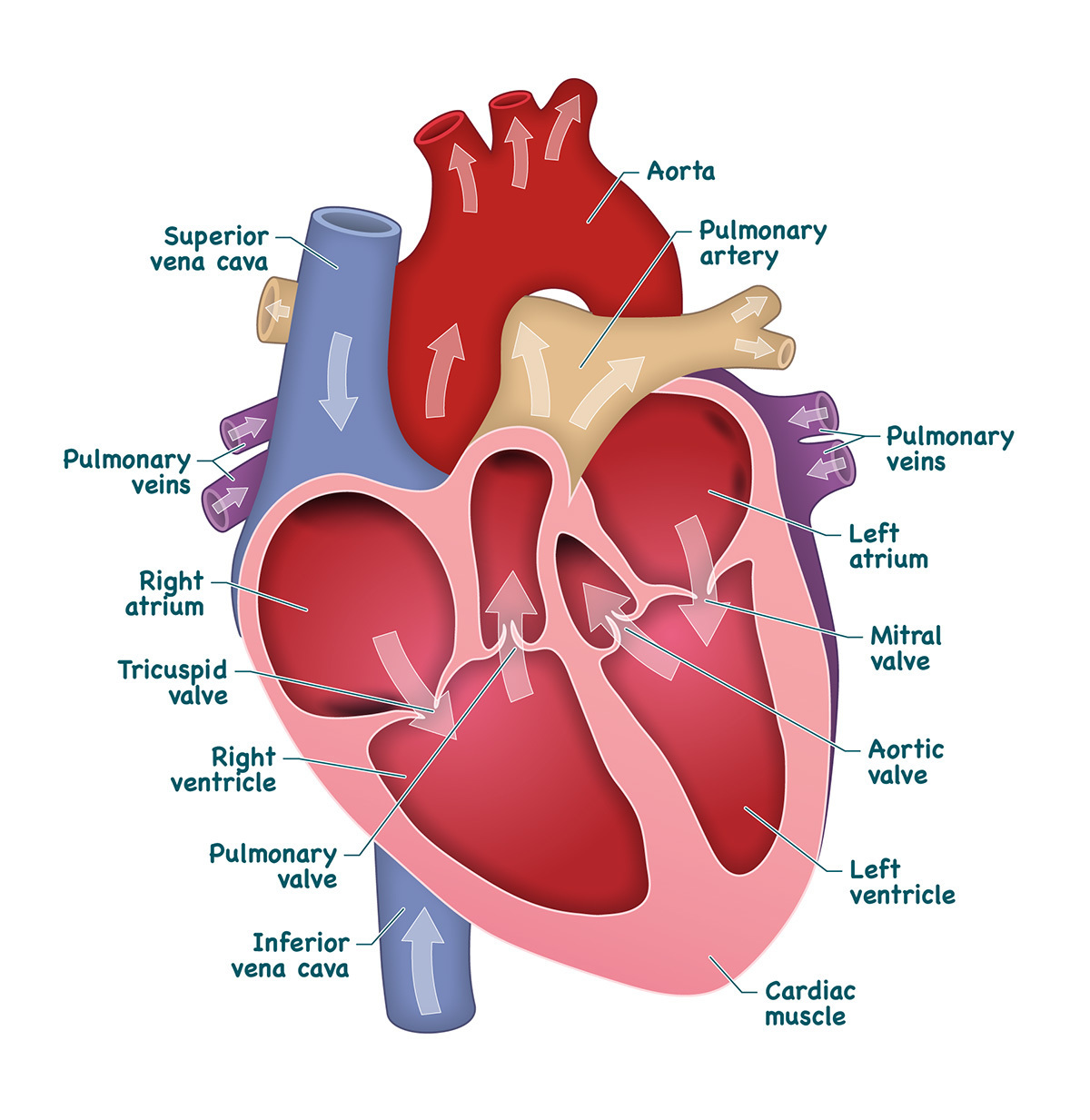
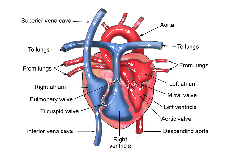
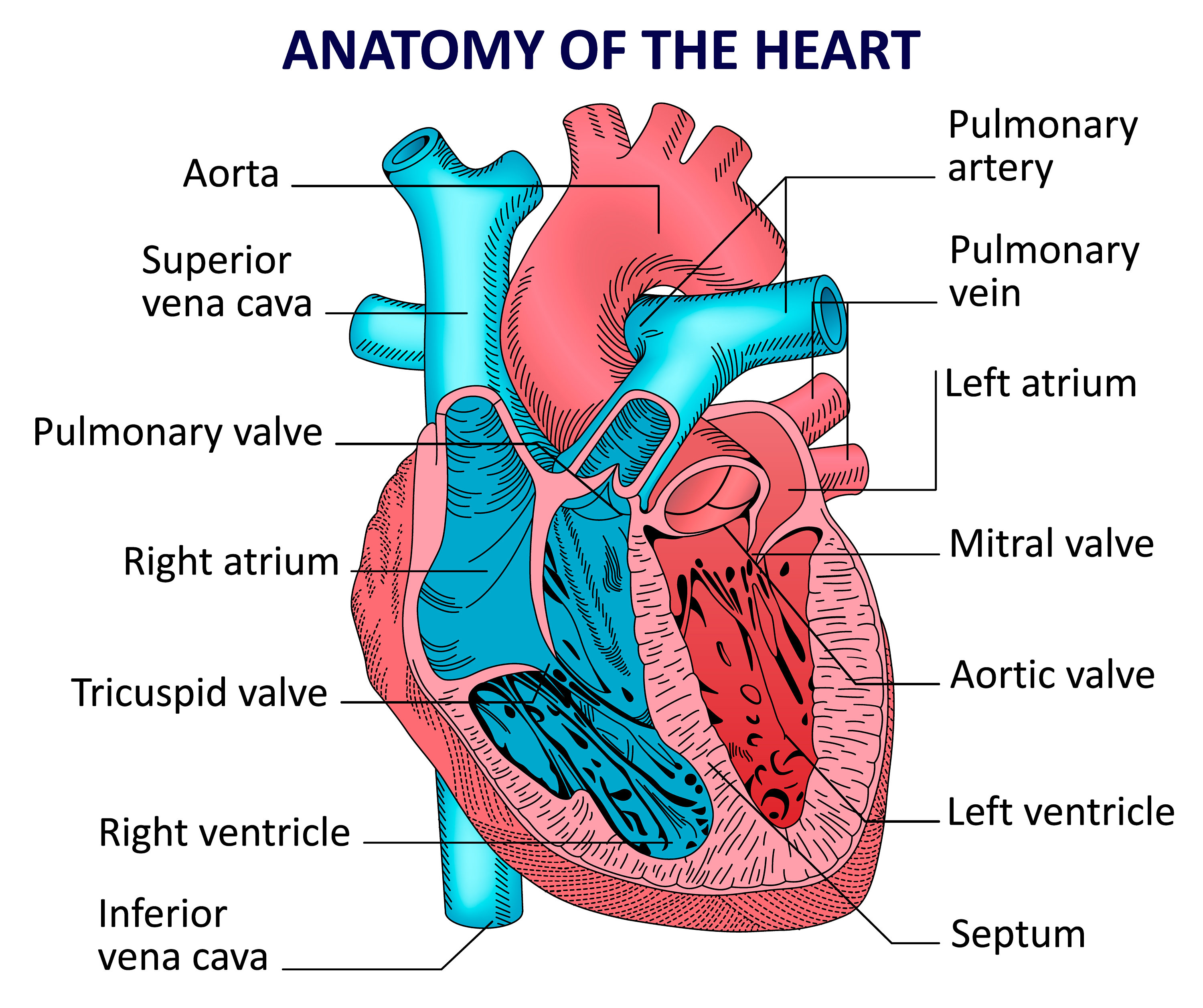
Labelled Heart Drawing - Web in this lecture, dr mike shows the two best ways to draw and label the heart! [right atrium and ventricle of the heart (labeled)] Images are labelled, providing an invaluable medical and anatomical tool. The heart is a hollow, muscular organ that pumps oxygenated blood throughout the body and deoxygenated blood to the lungs. It has four hollow chambers surrounded by muscle and other heart tissue. Electrical impulses make your heart beat, moving blood through these chambers. For optimal viewing of this interactive, view at your screen’s default zoom setting (100%) and with your browser window view maximised. Relate the structure of the heart to its function as a pump. Learn all about the heart, blood vessels, and composition of blood itself with our 3d models and explanations of cardiovascular system anatomy and physiology. See the labelling the heart activity for additional support in using this interactive. Web © 2024 visible body. By the end of this post, you will have a strong understanding of the main cardiac structures. Web how to draw the internal structure of the heart. The four types of valves are: Use a pen or pencil to draw the heart's main body. Describe the location and position of the heart within the body cavity. Your heart is in the center of your chest, near your lungs. This tool provides access to several medical illustrations, allowing the user to interactively discover heart anatomy. In addition to reviewing the human heart anatomy, we will also discuss the function and order in which blood flows. Web labeled heart diagrams. Web label the urinary tract #1 printout. Two atria (right and left) and two ventricles (right and left). Web the human heart is primarily comprised of four chambers. The right and left sides of the heart are separated by a muscle called the “septum.”. The upper two chambers of the heart are called auricles. The heart features four types of valves which regulate the flow of blood through the heart. The heart is a mostly hollow, muscular organ composed of cardiac muscles and connective tissue that acts. Web muscle and tissue make up this powerhouse organ. Web inside, the heart is divided into four. The heart is made up of four chambers: Web in this activity, students use online and paper resources to identify and label the main parts of the heart. Images are labelled, providing an invaluable medical and anatomical tool. Learn all about the heart, blood vessels, and composition of blood itself with our 3d models and explanations of cardiovascular system anatomy. Web anatomy of the human heart and coronaries: Your heart contains four muscular sections ( chambers) that briefly hold blood before moving it. The heart is made up of four chambers: Identify the tissue layers of the heart. Both sides work together to efficiently circulate the blood. Both sides work together to efficiently circulate the blood. Angle the slightly tampered end of the shape to the left about 120 degrees. Electrical impulses make your heart beat, moving blood through these chambers. The four types of valves are: Web label the urinary tract #1 printout. Web the heart is located in the thoracic cavity medial to the lungs and posterior to the sternum. Both sides work together to efficiently circulate the blood. It has four hollow chambers surrounded by muscle and other heart tissue. Web in just a few minutes, you will be able to label the entire diagram shown below! The video above also. The heart is a hollow, muscular organ that pumps oxygenated blood throughout the body and deoxygenated blood to the lungs. Web in this lecture, dr mike shows the two best ways to draw and label the heart! Rotate the 3d model to see how the heart's valves control blood flow between heart chambers and blood flow out of the heart.. 1.3m views 3 years ago 3 products. Two atria (right and left) and two ventricles (right and left). Your heart is in the center of your chest, near your lungs. They permit blood flow in one direction only, and prevent backflow of blood. The heart features four types of valves which regulate the flow of blood through the heart. The two upper chambers are called the atria, the remaining two lower chambers are the ventricles. The chambers are separated by heart valves, which make sure that the blood keeps flowing in the right direction. Both sides work together to efficiently circulate the blood. On its superior end, the base of the heart is attached to the aorta,mycontentbreak pulmonary arteries and veins, and the vena cava. But in a relief for the prime minister, the. The heart features four types of valves which regulate the flow of blood through the heart. Identify the main parts of a heart. Labeled heart diagram showing the heart from anterior. Web the human heart is primarily comprised of four chambers. By the end of this activity, students should be able to: Web in this activity, students use online and paper resources to identify and label the main parts of the heart. Web the heart is located in the thoracic cavity medial to the lungs and posterior to the sternum. Web we will use labeled diagrams and pictures to learn the main cardiac structures and related vascular system. Electrical impulses make your heart beat, moving blood through these chambers. Once you’re feeling confident, you can test yourself using the unlabeled diagrams of the parts of the heart below. They permit blood flow in one direction only, and prevent backflow of blood.Heart Anatomy Labeled Diagram, Structures, Blood Flow, Function of

heart PMG Biology
Heart And Labels Drawing at GetDrawings Free download
Human Heart Anatomy and Functions Location and Chambers
Heart And Labels Drawing at GetDrawings Free download
Revision notes of heart structure and labelled diagram
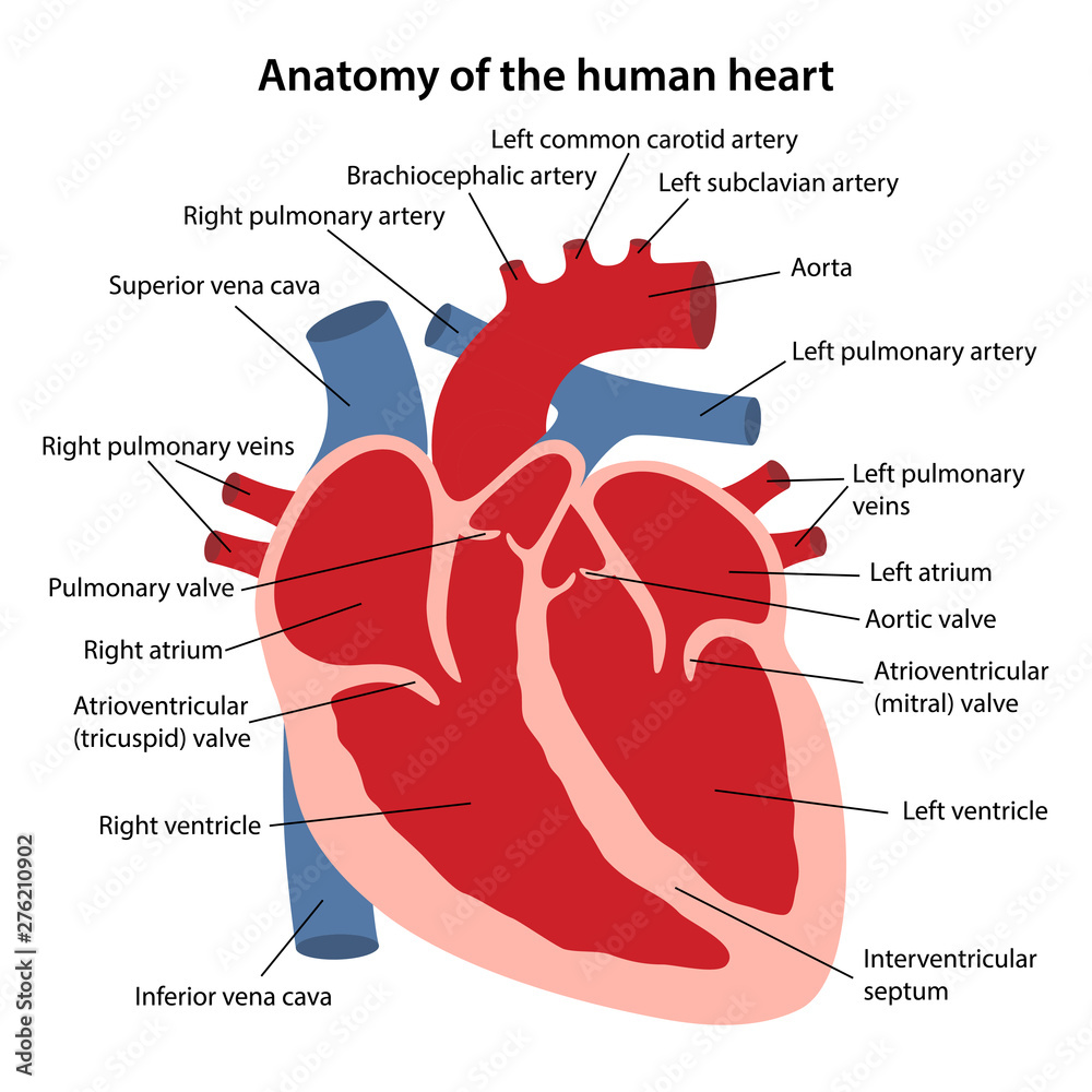
Anatomy of the human heart. Cross sectional diagram of the heart with

Labelled Heart
Human heart anatomy. Vector diagram Etsy

How to Draw the Internal Structure of the Heart 14 Steps
Take A Look At Our Labeled Heart Diagrams (See Below) To Get An Overview Of All Of The Parts Of The Heart.
The Inferior Tip Of The Heart, Known As The Apex, Rests Just Superior To The Diaphragm.
Web Heart Pictures, Diagram & Anatomy | Body Maps.
Web How To Draw The Internal Structure Of The Heart.
Related Post:
