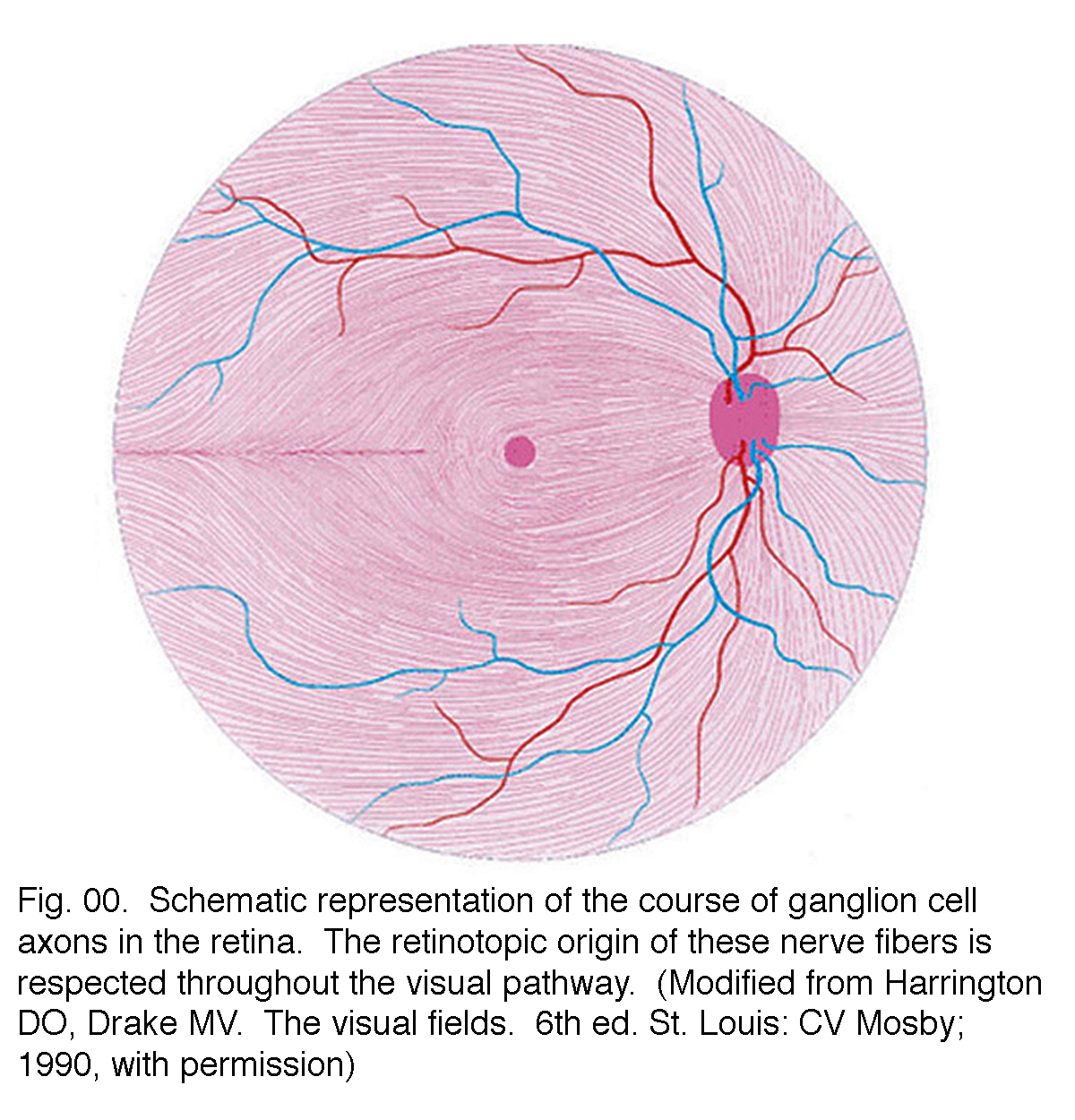
Retinal Drawing
Retinal Drawing - Web retinal drawings are a valuable tool allowing for easy visual communication of ophthalmoscopic findings despite less frequent use today in the era of digital medical records and abundance of imaging options (schachat et al. Can be used for serial follow up of patients to document changes in the pathology. Next, draw the macula temporal to it. The official description of cpt code 92201 is: Web most retinal surgeons are trained to create formal retinal drawings of the fundus. Web unique to retinal drawing, the inside of the globe is shown as circular images, forcing each artist to depict distortions tinted by their own interpretation. Code 92201 requires scleral depression, and your chart documentation should say so. First, trace the optic nerve on the retinal drawing. Drawings should include sufficient detail, standard colors and appropriate labels. The drawing must be separate and distinct from the comprehensive eye exam. (see a great example here ). The procedure pays about $27 each time, so mistakes can add up. Web most retinal surgeons are trained to create formal retinal drawings of the fundus. Acquired immunodeficiency syndrome (aids), cidofovir, cytomegalovirus (cmv), cmv retinitis fomivirsen, foscarnet, ganciclovir, ganciclovir implant, highly active antiretroviral therapy (hart), human immunodeficiency virus (hiv), immune recovery uveitis, valganciclovir. First,. Drawings should include sufficient detail, standard colors and appropriate labels. Web retinal drawings must be maintained in the patient’s record. Web drawing of the retina in the left eye: Draw it on the right for the right eye, and on the left for the left eye. Aetna considers extended ophthalmoscopy with a detailed retinal drawing for evaluation of the posterior. 91 views 1 year ago. Retinal drawing must be maintained in the patient’s record. The correct code for this eo would be 92225 ophthalmoscopy, extended, initial. Web unique to retinal drawing, the inside of the globe is shown as circular images, forcing each artist to depict distortions tinted by their own interpretation. The drawing must be separate and distinct from. Drawings should include sufficient detail, standard colors and appropriate labels. Individual drawings should be made for each eye. Web a true retina drawing will contain three concentric circles. Acquired immunodeficiency syndrome (aids), cidofovir, cytomegalovirus (cmv), cmv retinitis fomivirsen, foscarnet, ganciclovir, ganciclovir implant, highly active antiretroviral therapy (hart), human immunodeficiency virus (hiv), immune recovery uveitis, valganciclovir. Code 92201 requires scleral depression,. There should also be 12 tick marks indicating each clock hour of the retina. It is a useful reference to monitor the clinical process and also at the time of surgery. The first represents the equator, the second represents the ora serrata, and the third represents the pars plana. Drawings should include sufficient detail, standard colors and appropriate labels. Lets. Web the two replacement codes are defined as follows: This colorful book examines the technique and history of retinal drawing, its application and variations of artistic rendition, and covers a wide array of visually distinctive disorders. Lets take a look into the most important colors for retinal drawing. A lost art of medicine | find, read and cite all the. Aetna considers extended ophthalmoscopy with a detailed retinal drawing for evaluation of the posterior portion of the eye following routine ophthalmoscopy medically necessary for any of the following indications: A lost art of medicine | find, read and cite all the research you need on researchgate. The official description of cpt code 92201 is: The procedure pays about $27 each. Next, draw the macula temporal to it. Aetna considers extended ophthalmoscopy with a detailed retinal drawing for evaluation of the posterior portion of the eye following routine ophthalmoscopy medically necessary for any of the following indications: There should also be 12 tick marks indicating each clock hour of the retina. Lets take a look into the most important colors for. Web a detailed retinal drawing is required. (see a great example here ). Code 92201 requires scleral depression, and your chart documentation should say so. The drawing must be separate and distinct from the comprehensive eye exam. There should also be 12 tick marks indicating each clock hour of the retina. Pdf | on jul 1, 2011, luann dvorak and others published retinal drawing: With retinal drawing and scleral depression of peripheral retinal disease (e.g., for retinal tear, retinal detachment, retinal tumor) with interpretation and report, unilateral or bilateral. Aetna considers extended ophthalmoscopy with a detailed retinal drawing for evaluation of the posterior portion of the eye following routine ophthalmoscopy medically. The correct code for this eo would be 92225 ophthalmoscopy, extended, initial. Horseshoe shapes at 1:30 with accompanying ablatio retinae from 1 to 3 o’clock and degeneration area at 10:30 with a small round hole. Web a true retina drawing will contain three concentric circles. Although payer policies differ, some common charting requirements include: Draw it on the right for the right eye, and on the left for the left eye. Pdf | on jul 1, 2011, luann dvorak and others published retinal drawing: (see a great example here ). Web most retinal surgeons are trained to create formal retinal drawings of the fundus. Web a detailed retinal drawing is required. The official description of cpt code 92201 is: The first represents the equator, the second represents the ora serrata, and the third represents the pars plana. Web because cpt code 92201 reports peripheral retinal drawings, you can expect the list of valid indications to include conditions of the peripheral retina and vessels rather than disease in the center of the fundus or near the optic nerve. With retinal drawing and scleral depression of peripheral retinal disease (eg, for retinal tear, retinal detachment, retinal tumor) with interpretation and report, unilateral or bilateral.’. The drawing must be separate and distinct from the comprehensive eye exam. Web drawing of the retina in the left eye: Web retinal drawings are a valuable tool allowing for easy visual communication of ophthalmoscopic findings despite less frequent use today in the era of digital medical records and abundance of imaging options (schachat et al.
Gross Anatomy Of Retina ANATOMY
Simple Anatomy of the Retina by Helga Kolb Webvision
Retinal Drawing at GetDrawings Free download

ME120.751 Ophthalmological Illustration Art as Applied to Medicine

Types of Retinal Disorders San Diego, CA

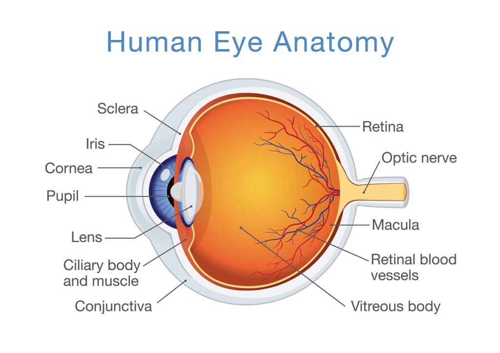
Human eye anatomy, retina detailed illustration. Human eye anatomy

The basic retinal structure. Histological appearance of choroid and
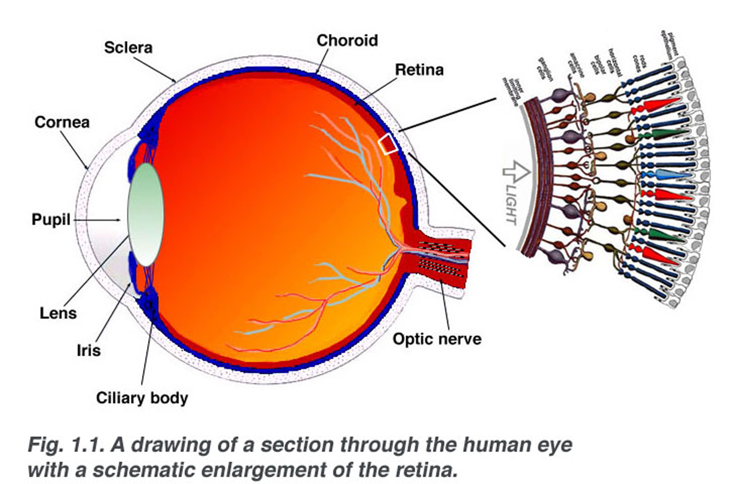
Simple Anatomy of the Retina by Helga Kolb Webvision
Basic Anatomy of Retina Elman Retina Group Eye Doctors
Retinal Drawing at GetDrawings Free download
Individual Drawings Should Be Made For Each Eye.
Web The Retina Specialist Confirms The Diagnosis Of Macular Hole In The Right Eye And Documents It With A Retinal Drawing With Labels And An Interpretation And Report.
This Colorful Book Examines The Technique And History Of Retinal Drawing, Its Application And Variations Of Artistic Rendition, And Covers A Wide Array Of Visually Distinctive Disorders.
Acquired Immunodeficiency Syndrome (Aids), Cidofovir, Cytomegalovirus (Cmv), Cmv Retinitis Fomivirsen, Foscarnet, Ganciclovir, Ganciclovir Implant, Highly Active Antiretroviral Therapy (Hart), Human Immunodeficiency Virus (Hiv), Immune Recovery Uveitis, Valganciclovir.
Related Post: